Different groups reported using Indo as multiphoton
fluorescence marker with excitation between 690 nm and 720 nm.
References:
Jones, K.T., Soeller, C. & Cannell, M.B. (1998) The passage of Ca2+ and
fluorescent markers between the sperm and egg after fusion in the mouse.
Development 125, 4627-4635.
Del Mar Photonics supplies multi-photon lasers and systems based on cost
effective femtosecond sources:
Multiphoton Imaging
Related Del Mar Photonics products
If you can't find information about the products that
you are looking for just
e-mail our Sales Team and we'll e-mail or call you back right away!
Dear friends,
I am proud to inform that our short review on multiphoton microscopy is the most
read paper on biomedical engineering online:
http://www.biomedical-engineering-online.com/content/5/1/36.
All the best
Alby
ISTITUTO ITALIANO
DI TECNOLOGIA
Prof. Alberto Diaspro
Scientific Head
Nanophysics
Via Morego, 30 16163 Genova
Tel: +39-010.71.781.503
Fax +39-010-72.03.21
Mobile +39-3666719968
www.iit.it
alberto.diaspro@iit.it
Multi-photon excitation microscopy Alberto Diaspro1,2,5 , Paolo Bianchini1 , Giuseppe Vicidomini1 ,
Mario Faretta3 , Paola Ramoino4 and Cesare Usai5
1 LAMBS-MicroScoBio Research Center, Department of Physics, University of Genoa,
Via Dodecaneso 33, 16146 Genova, Italy
2 IFOM The FIRC Institute for Molecular Oncology Foundation, Via Adamello, 16,
20139 Milan, Italy
3 IFOM-IEO Consortium for Oncogenomics European Institute of Oncology, via
Ripamonti 435, 20141 Milan, Italy
4 DIPTERIS – Department for the Study of the Territory and its Resources,
University of Genoa, Corso Europa 26, 16132 Genova, Italy
5 CNR- National Research Council, Institute of Biophysics, Via De Marini, 6,
16149 Genova, Italy
author email corresponding author email
Multi-photon excitation (MPE) microscopy plays a growing role among
microscopical techniques utilized for studying biological matter. In conjunction
with confocal microscopy it can be considered the imaging workhorse of life
science laboratories. Its roots can be found in a fundamental work written by
Maria Goeppert Mayer more than 70 years ago. Nowadays, 2PE and MPE microscopes
are expected to increase their impact in areas such biotechnology, neurobiology,
embryology, tissue engineering, materials science where imaging can be coupled
to the possibility of using the microscopes in an active way, too. As well, 2PE
implementations in noninvasive optical bioscopy or laser-based treatments point
out to the relevance in clinical applications. Here we report about some basic
aspects related to the phenomenon, implications in three-dimensional imaging
microscopy, practical aspects related to design and realization of MPE
microscopes, and we only give a list of potential applications and variations on
the theme in order to offer a starting point for advancing new applications and
developments.
1. Introduction
There have been a variety of reasons for the continuing growth of interest in
optical microscopy in spite of the low resolution with respect to modern
scanning probe or electron microscopy [1]. The main reason lies in the fact that
optical microscopy is still considered unique in allowing the 4D (x-y-z-t)
examination of biological systems in a hydrated state in living samples or under
experimental conditions that are very close to living or physiological states.
This evidence coupled to fluorescence labelling and other advances in molecular
biology permits to attack in an effective way the complex and delicate problem
of the connection between structure and function in biological systems [2-6].
Within this framework, inventions in microscopy were stimulated, and contributed
to the evolution of the optical microscope in its modern forms [7,8].
Multiphoton excitation microscopy is an important part of this progress in the
field of microscopy applied to the study of biological matter from the
inventions of the confocal microscope [9] and of the atomic force microscope
[10]. Nowadays, confocal and multiphoton microscopes can be considered the
imaging workhorses of life science laboratories [11].
Multiphoton excitation microscopy (MPE) has its roots in two-photon excitation
(2PE) microscopy whose story dates back more than 70 years. In 1931, Maria
Goeppert-Mayer published her brilliant doctoral dissertation on the theory of
two-photon quantum transitions in atoms and established the theoretical basis
behind 2PE [12]. This photophysical effect was experienced after the development
of laser sources, as well, other non-linear related effects were also observed
in the 60s and 70s [13,14]. In 1976, Berns reported about a probable two-photons
effect as a result of focusing an intense pulsed laser beam onto chromosomes of
living cells [15], and such interactions form the basis of modern nanosurgery
[16] and targeted transfection [17]. One has to wait until 1978 to find the
description of the first nonlinear scanning optical microscope with depth
resolution. The reported microscopic imaging was based on second-harmonic
generation (SHG) and the possibility of performing 2PE microscopy was outlined
[18]. Unfortunately, applications in biology were hampered due by the high peak
intensities required for priming 2PE fluorescence. This obstacle was overcome
with the advent of ultrashort and fast pulsed lasers in the 80s [19]. Denk and
colleagues in a seminal paper on 2PE laser scanning fluorescence microscopy
clearly demonstrated the capability of 2PE microscopy for biology [20]. This
fact brought a "new deal" in fluorescence microscopy [21-23]. Such a leap in
scientific technology stimulated disparate disciplines and several variations on
the theme extending studies from the tracking of individual molecules within
living cells to the observation of whole organisms [24-28].
2. Foundations of 2PE microscopy
Two-photon excitation of fluorescent molecules is a non-linear process related
to the simultaneous absorption of two photons whose total energy equals the
energy required for the more familiar one-photon excitation (1PE) [29]. The
excitation process of a fluorescent molecule under 1PE typically requires
photons in the ultraviolet or blue/green spectral range. Under sufficiently
intense illumination, usually provided by a laser source, the very same process,
i.e. excitation of a fluorescent molecule from the ground to the excited state,
can take place in the infrared spectral range. This situation is illustrated in
figure 1 using a Perrin-Jablonski diagram: the sum of the energies of the "two"
infrared photons colliding with the very same molecule has to be greater than
the energy gap between the molecule's ground and excited states. Since 2PE
excitation requires at least two statistically "independent" photons for each
excitation process, its rate depends on the square power of the instantaneous
intensity. 3PE and higher photon excitation is also possible. This implies that
deep ultraviolet (UV) microscopy can be performed without having the
disadvantages related to UV-matter interactions, i.e. polymerization or
temperature effects. However, our treatment will be mainly conducted in terms of
2PE for sake of simplicity but any step can be extended to MPE.
Figure 1. Perrin-Jablonski fluorescence diagram. Simplified Perrin-Jablonski
scheme for 1PE, 2PE and 3PE. Once the excited state has been reached the
subsequent fluorescent emission is the very same for the three different
modalities of excitation.
The most popular relationship about 2PE is related to the practical situation of
a train of beam pulses focused through a high numerical aperture (NA) objective,
with a duration τp and fprepetition rate. The advantage of using a train of
repeated beam pulses is given by the fact that high peak power can be used for
priming the process resulting on an average power tolerated by the biological
system under investigation. Under controlled conditions, the probability, na,
that a certain fluorophore simultaneously absorbs two photons during a single
pulse, in the paraxial approximation is given by [20]
where Pave is the average power of the illumination beam, δ2 is the two-photon
cross section of the fluorescent molecule, and λ is the excitation wavelength.
Introducing 1 GM (Goppert-Mayer) = 10-58 [m4 s], for a δ2 of approximately 10
GM, focusing through an objective of NA >1, an average incident laser power of ≈
1–50 mW, operating at a wavelength ranging from 680 to 1100 nm with 80–150 fs
pulsewidth and 80–100 MHz repetition rate, one should get fluorescence without
saturation. It is convenient that for optimal fluorescence generation, the
desirable repetition time of pulses should be on the order of typical
excited-state lifetime, which is a few nanoseconds for commonly used fluorescent
molecules. For this reason the typical repetition rate is around 100 MHz, i.e.
one order of magnitude slower than typical fluorescence lifetime. As well,
during the pulse time (10-13 s of duration and a typical lifetime in the 10-9 s
range) the molecule has insufficient time to relax to the ground state. This can
be considered a prerequisite for absorption of another photon pair. Therefore,
whenever na approaches unity saturation effects start occurring. Such a
consideration allows one to optimize optical and laser parameters in order to
maximize the excitation efficiency without saturation. In case of saturation the
resolution is declining and worsening the image. It is also evident that the
optical parameter for enhancing the process in the focal plane is the lens
numerical aperture, NA, even if the total fluorescence emitted is independent
from this parameter. This value is usually kept around 1.3–1.4. As example, for
fluorescein that possesses a δ2 ≅38 GM at 780 nm, using NA = 1.4, a repetition
rate of 100 MHz and pulse width of 100 fs within a range of Pave assumed 1, 10,
20 and 50 mW, from equation (1) one has na≈ 5930 (Pave)2. This means that, as
function of the average excitation power 1, 10, 20, 50 mw one gets 5.93 10-3,
5.93 10-1, 1.86, 2.965 respectively, with saturation starting at 10 mW. The
related rate of photon emission per molecule, at a non saturation excitation
level, in absence of photobleaching, is given by na multiplied by the repetition
rate of the pulses, i.e. approximately 5·107 photons s-1. It is worth noting
that when considering the effective fluorescence emission one should consider a
further factor given by the so-called quantum efficiency of the fluorescent
molecules. It is worth noting that the fluorophore emission spectrum results
independent of the excitation mode from 1PE to MPE like the quantum efficiency.
Now, even if the quantum-mechanical selection rules for MPE differ from those
for one-photon excitation, several common fluorescent molecules can be used.
Unfortunately, the knowledge of 1PE cross-section for a specific fluorescent
molecule does not allow any quantitative prediction of the two-photon trend. The
only "rule of thumb" that one can use states that a 2PE cross-section peak can
be expected at a 2 folds wavelength with respect to the 1PE case. Table 1
summarises the excitation properties of some popular fluorescent molecules under
2PE regime.
Table 1. 2PE excitation parameters.
3. Optical implications of 2PE
3.1 Optical sectioning and confocal imaging
The possibility of the three-dimensional reconstruction of the volume
distribution of intensive parameters, as fluorescence emission, from biological
systems is one of the most powerful properties of the optical microscope. To
collect optical slices from a three-dimensional object the so-called optical
sectioning [3,11] technique is used as depicted in figure 2. It is essentially
based on a fine z stepping either of the objective or of the sample stage,
coupled with the usual x-y image capturing. The synchronous x-y-z scanning
allows the collection of a set of two-dimensional images, which are somehow
affected by signal cross talk from other planes from the sample. In fact, the
observed image Oj, obtainable when positioning the geometrical focus of the lens
at a certain plane" j "within the specimen, is produced by the true fluorescence
distribution Ij at plane "j", distorted by the microscope in some way that can
be described by a function S, plus differently distorted contributions from
adjacent "k" planes positioned above and below the actual plane, and noise N.
Using a convenient and appropriate formalism one has:
Figure 2. Optical sectioning scheme. A three-dimensional sample can be sketched
as a series of optical slices. Let's call slice j the one containing the
geometrical focus of the objective and refer to the adjacent planes as k slices.
The sample contains a three-dimensional distribution of fluorescently labelled
molecules whose intensity distribution is I, slice by slice. The thickness of
each optical slice is approximately one half of the axial resolution, say ≈λ/2.
Oj = IjSj + ∑k≠j IkSk + N. (2)
Equation (2) reflects the fact that when a set of two-dimensional images is
acquired at various focus position and under certain conditions, in principle,
one can recover the 3D shape of the object, described by the intensive parameter
I, by solving the above set of equations and finding the best estimate for I,
slice by slice. By this procedure, unwanted light can be computationally removed
combining the image data from a stack of "k" images. Such an operation can be
optically performed using some physical stratagems that are behind confocal and
MPE/2PE scanning microscopy.
In confocal microscopy, the observed image is built up, scanning the sample
point by point, by adding information from x-y-z sampled regions. The price to
pay, or the main drawback, is that image formation is not as immediate as
widefield techniques in which the whole image is acquired at the same time. This
is due to the fact that in confocal microscopy the specimen is sequentially
illuminated point by point and at the same time all is masked but the
illuminated in-focus regions for providing return light to the detector. As
shown in figure 3 an illumination and a detection pinhole are placed in the
optical pathway. The detection pinhole – the mask – is placed in front of the
detector at a plane that is conjugate to the in-focus or "j" plane, such that
the illumination spot and the pinhole aperture are simultaneously focused at the
same specimen volume. This coincidence of the illumination and detected volume
is responsible for confocality. The final effect of such an optical
implementation is that out-of-focus contributions are excluded from the detector
surface and the observed image, Oj, is close to the true distribution of the
fluorescence intensity I, plane by plane during the x-y-z scanning operations.
This allows performing optical sectioning.
Figure 3. Confocal optical pathways. An illumination and a detection pinhole are
placed in the optical pathway. The detection pinhole – the mask – is placed in
front of the detector at a plane that is conjugate to the in-focus or "j" plane,
such that the illumination spot and the pinhole aperture are simultaneously
focused at the same specimen volume. This coincidence of the illumination and
detected volume is responsible for confocality. The illumination pinhole allows
to perform pointlike scanning.
3.2 The 2PE optical case
In terms of optical implications the two-photon effect has the important
consequence of limiting the excitation region to within a sub-femtoliter volume.
This means that the emission region is intrinsically confocal. The resulting 3D
confinement in terms of image formation process can be described by means of
consolidated optical considerations [30]. Using a certain excitation light at a
wavelength λ, the intensity distribution within the focal region of an objective
having numerical aperture NA = n sin (α) is given, in the paraxial regime, by
where J0 is the 0th order Bessel function, ρ is a radial coordinate in the pupil
plane, n is the refractive index of the medium between the lens and the
specimen, (α) is the semi-angle of aperture of the lens [31],
and
are dimensionless axial and radial coordinates, respectively, normalized to the
wavelength. Now, the intensity of fluorescence distribution within the focal
region has a I(u, v) behaviour for the 1PE case [31]. In case of 2PE one has to
consider a double wavelength and a square behaviour, i.e. I2(u/2, v/2). As
compared with the 1PE case, the 2PE emission intensity distribution is axially
confined.
In fact, considering the integral over ν, keeping u constant, its behaviour is
constant along z for one-photon and has a half-bell shape for 2PE. This
behaviour is responsible of the 3D discrimination property of 2PE, i.e. of the
optical sectioning properties of the 2PE microscope.
Now, the most interesting aspect is that the excitation power falls off as the
square of the distance from the lens focal point, within the approximation of a
conical illumination geometry [31]. In practice this means that the square
relationship between the excitation power and the fluorescence intensity brings
about the fact that 2PE falls off as the fourth power of distance from the focal
point of the objective. This fact implies that those molecules away from the
focal region of the objective lens do not contribute to the image formation
process and are not affected by photobleaching or phototoxicity. Since these
molecules are not involved in the excitation process, a confocal-like effect is
obtained without the necessity of a confocal pinhole. It is immediately evident
that in this case the optical sectioning effect is obtained in a physically
different way with respect to the confocal case. Accordingly the optical set-up
is simplified, under some aspects, see figure 4.
Figure 4. MPE simplified optical pathways. In the MPE optical pathways the
emission pinhole is removed since the only emitted light reaching the sensor is
coming from the currently point scanned volume in the sample. No other
fluorescence signal is produced elsewhere.
Figure 5 and figure 6 show the differences in terms of excitation-emission
process between confocal and multiphoton schemes, respectively. The consequences
of the spatial confinement of the MPE result in a consequent confinement of the
emitted fluorescence: in the confocal case all the molecules within the double
cone of excitation are involved in the light-matter interaction while in the MPE
case such interaction is restricted to a small volume centred at the geometrical
focus of the objective. The immediate consequence is that a 2PE microscope is an
intrinsically three-dimensional image formation system. This fact has also very
important consequences on the photobleaching processes. So far, in the 2PE case
no fluorescence has to be removed from the detection pathway since fluorescence
can exclusively come from a small focal volume that has a capacity of the order
of fraction of femtoliter. In fact, in 2PE over 80% of the total intensity of
fluorescence comes from a 700–1000 nm thick region about the focal point for
objectives with numerical apertures in the range from 1.2 to 1.4 [30]. The fact
that the background signal coming from adjacent planes tends to zero produces a
sort of compensation for the reduced spatial resolution due to the utilization
of a wavelength that is twice with respect to the 1PE case, as shown in figure
7. On the other hand, the utilisation of infrared wavelengths instead of
UV-visible ones allows achieving a deeper penetration than in conventional case
[32]. This is due to the fact that the scattering effect is proportional to the
inverse fourth power of the wavelength. Thus the longer wavelengths used in 2PE,
or in general in MPE, will be scattered less than the ultraviolet-visible
wavelengths used for conventional excitation allowing to reach fluorescence
targets in depth within thick samples (approx. 1 mm). It has been shown that
two-photon fluorescence images can be obtained throughout almost the entire grey
matter of the mouse neocortex by using optically amplified femtosecond pulses.
The achieved imaging depth approaches the theoretical limit set by excitation of
out-of-focus fluorescence [33]. The fluorescence light emitted, on the way back,
can be more efficiently collected using a large area detector since it can
uniquely come from the sub-femtoliter 2PE volume of event.
Figure 5. Confocal fluorescence emission distribution. The emission process, in
green, under blue 1PE excitation is broadened in the whole double cone
excitation shape within the analyzed cell.
Figure 6. MPE fluorescence emission distribution. Confinement of the emission
process, in green, under red 2PE. Under 2PE the only molecules excited are those
confined in a small subfemtoliter volume at the illumination beam focus
position. This is particularly relevant for the photobleaching process that is
globally reduced with respect to the 1PE case. The capacity of the volume can be
approximatively calculated by using the resolution parameters of the optical
system, since they are indicators of the volume containing the maximum photon
density. This is valid only for non saturated processes. Under saturation of
fluorescence beam intensity plays an important role in the emission shape and
optical resolution.
Figure 7. Pointlike emitter optical response. From left to right: calculated x-y
(above) and r-z (below) intensity distributions, in logarithmic scale, for a
point like source imaged by means of wide-field, 2PE and confocal microscopy.
Both 2PE and confocal shapes exhibit a better signal to noise ratio than
widefield case. 2PE distribution is larger due the fact that a wavelength twice
than in the wide-field and confocal case is responsible for the intensity
distribution. Such intensity distributions are also known as point spread
functions of the related microscopes. Optical conditions: excitation wavelengths
are 488 nm and 900 nm for 1PE and 2PE, respectively; emission wavelength is 520
nm; numerical aperture is 1.3 for an oil immersion objective with oil refractive
index value set at 1.515.
4. Practical aspects for the realization of a 2PE microscope
The main elements to realize a 2PE/MPE architecture, including confocal
modality, are the following: high peak-power laser delivering moderate average
power (fs or ps pulsed at relatively high repetition rate) emitting infrared or
near infrared wavelengths (650–1100 nm), laser sources for confocal 1PE, a laser
beam scanning system, high numerical aperture objectives (>1), a high-throughput
microscope pathway, a spectral separation module for the emitted signal
discrimination, and a high-sensitivity detection system [34]. Figure 8 shows a
general scheme for a MPE microscope also illustrating two popular approaches
that can be used for image formation, namely: de-scanned and non de-scanned
mode. The former uses the very same optical pathway and mechanism employed in
confocal laser scanning microscopy. The latter mainly optimises the optical
pathway by minimising the number of optical elements encountered on the way from
the sample to detectors, and increases the detector area. MPE non-descanned mode
allows very good performances providing superior signal-to-noise ratio inside
strongly scattering samples [32,33]. In the de-scanned approach pinholes are
removed or set to their maximum aperture and the emission signal is captured
using the very same optical scanning pathway used for excitation. In the latter,
the aim is to optimize the collection efficiency: pinholes are removed and the
radiation emitted without passing through the laser beam scanning mirrors.
Photomultiplier tubes are the most popular detectors in MPE microscopy.
Avalanche photodiodes are also excellent in terms of sensitivity exhibiting
quantum efficiency close to 70%–80% in the visible spectral range. Unfortunately
they are high cost and the small active photosensitive area could introduce
drawbacks in the detection scheme and require special de-scanning optics. CCD
cameras are generally used in video rate multifocal imaging. Laser sources
represent the core element for the 2PE/MPE microscope since for MPE high photon
flux densities are required, > 1024 photons cm-2s-1. Using radiation in the
spectral range of 650–1100 nm for MPE, excitation intensities in the MW-GW cm-2
have to be produced. Nowadays, laser sources suitable for 2PE can be described
as "turnkey" systems, and Ti Sapphire lasers are the most utilized due to the
high coincidence with the 2PE wavelengths needed for the majority of the
commonly used fluorescent molecules. Other laser sources used for 2PE are
Cr-LiSAF, pulse-compressed Nd-YLF in the femtosecond regime, and mode-locked
Nd-YAG and picosecond Ti-Sapphire lasers in picosecond regime. Moreover the
absorption coefficients of most biological samples, cells and tissues are
minimised within this spectral window. Table 2 reports data on the currently
available laser sources for applications in MPE microscopy and spectroscopy. The
parameters that are more relevant in the selection of the laser source are
average power, pulsewidth and repetition rate, and wavelength also accordingly
to equation (1). The most popular features for an infrared pulsed laser are
700mW-1W average power, 80–100 MHz repetition rate, and 100–150 fs pulse width.
So far, the use of short pulses and high repetition rates are mandatory to allow
image acquisition in a reasonable time while using power levels that are
biologically tolerable. In order to minimise pulse width dispersion problems it
should be considered to operate with pulses around 150 nm. This constitutes a
very good compromise both for pulse stretching and sample viability. One should
always remind that a shorter pulse broadens more than a longer one. Advances in
laser sources are going on considering more compact sources, large tunability
range, high average power, and special designs for tailored needs at lower
prices [35]. Objective lenses influence the performances of any optical
microscope, and for a MPE system special considerations have to be done taking
into the proper account both equations (1) and (3). New technological requisites
have to be considered with respect to conventional excitation fluorescence
microscope. An adequate transmission in the IR regime has to be coupled with
good collection efficiency towards the ultraviolet region. Moreover, the number
of components should be minimised without affecting resolution properties in
order to reduce pulse widths distortions. Although the collection efficiency of
the time averaged photon flux is dependent on the numerical aperture of the
collecting lens, the total fluorescence generation is independent of the
numerical aperture of the focusing lens when imaging thick samples. Figure 9
shows an example of optical sectioning performed through a thick sample that
exploits the autofluorescence signal. This is due to the fact that the increase
of intensity, obtained by a sharper focusing (high NA), is counterbalanced by
the shrinking of the excitation volume. Thus the total amount of fluorescence
summed over the entire space remains constant. The very relevant practical
consequence of this fact is that in 2PE measurements on thick samples, assuming
no aberrations, the generated fluorescence is insensitive to the size of the
focal spot. As a positive consequence, a moderate variation of the laser beam
size would not affect the measurements. This is a very efficient condition due
to the fact that using an appropriate (non-descanned) acquisition scheme it is
possible to collect all the generated fluorescence. In terms of pulse
broadening, a 100 fs pulse can result between 1.14 and 1.23 times at the sample
using a good lens. Once a MPE architecture has been realized, one should
consider to keep under control the following parameters: power and pulse width
at the sample focal plane checking for the square intensity/power behaviour,
spectral separation of the emitted fluorescence including removal of the
possible excitation reflections that could be particularly subtle, z-axis
precise control and laser-scanning system alignment [34]. Figure 10 and figure
11 demonstrate the multiple fluorescence imaging capability and 3D multiple
fluorescence imaging, respectively.
Figure 8. MPE simplified optical schemes. Descanned (red dot) and non-descanned
(blue dot) schemes for 2PE microscopy. Due to the confinement of the excitation
process when operating in non-descanned mode the collection efficiency in depth
is increased as shown by the two side views from the very same thick scattering
sample (Courtesy of Mark Cannel and Christian Soeller; image inset courtesy of
Ammasi Periasamy).
Table 2. MPE laser table.
Figure 9. Optical sectioning using 2PE autofluorescence. 2PE optical sectioning
of Colpoda maupasi resting cysts, 21–32 μm average dimensions. Encystment is
particularly widespread in species living in ephemeral fresh-water puddles and
is induced by exhaustion of the food and drying out. The resulting images have
been obtained at LAMBS-MicroScoBio exploiting autofluorescence primed using 740
nm excitation. A Chameleon-XR ultrafast Ti-Sapphire laser (Coherent Inc., USA)
and a Nikon PCM2000 confocal scanning head have been used [34]. Linear frame
dimension is 70 μm, z steps have been performed every 0.5 μm, and not all the
optical sections are imaged.
Figure 10. Multiple fluorescence 2PE imaging. 2PE multiple fluorescence image
from a 16 μm cryostat section of mouse intestine stained with a combination of
fluorescent stains (F-24631, Molecular Probes). Alexa Fluor 350 wheat germ
agglutinin, a blue-fluorescent lectin, was used to stain the mucus of goblet
cells. The filamentous actin prevalent in the brush border was stained with
red-fluorescent Alexa Flu or 568 phalloidin. Finally, the nuclei were stained
with SYTOX ® Green nucleic acid stain. Imaging has been performed at 780 nm, 100
x 1.4 NA Leica objective, using a Chameleon XR ultrafast Ti-Sapphire laser
(Coherent Inc., USA) coupled at LAMBS-MicroScoBio with a Spectral Confocal Laser
Scanning Microscope, Leica SP2-AOBS.
Figure 11. 3D and 2D fluorescence projections. Pictorial representation of the
3D and 2D projections of multiple fluorescence from a marine sponge, Chondrilla
nucula. The specimen has been loaded with Alexa 488 fluorescent molecules
specific aminobutirric acid (GABA) emitting in green, DAPI for nuclear DNA for
the blu component. Red signals are due to the autofluorescence from symbiontic
bacteria contamination. Imaging has been perfomed using a Chameleon XR ultrafast
Ti-Sapphire laser (Coherent Inc., USA) coupled at LAMBS-MicroScoBio with a
Spectral Confocal Laser Scanning Microscope, Leica SP2-AOBS. (Sample
availability and preparation, courtesy of Renata Manconi, University of Sassari,
Roberto Pronzato and Lorenzo Gallus, University of Genoa).
5. Application trends and conclusions
2PE and MPE microscopes are expected to increase their impact in areas such
biotechnology, neurobiology, embryology, tissue engineering, materials science
where imaging can be coupled to the possibility of using the microscopes in an
active way, too. Clinically, 2PE may find applications in non-invasive optical
bioscopy, while in cell biology the imaging abilities are coupled to the
possibility of producing localized chemical reactions. Potential applications to
integrative cardiac physiology or the possibility of tracking for long time
biological events in living systems point out to the ability of making direct
observations of phenomena and circumstances that before could only be inferred
using other approaches. The myriad of new investigation possibilities offered by
2PE/MPE microscopy enlarges so much the fields of application that it is not
possible to outline in a complete way all the variations that can take place.
For this reason, we give in this last paragraph asummary of main properties of
MPE and a limited overview of paramount trends.
The great impact of 2PE in optical microscopy is related to the fact that it
couples a three-dimensional intrinsic ability with almost five other interesting
capabilities [21]. First, 2PE greatly reduces photo-interactions and allows
imaging of living specimens on long time periods. Second, it allows operating in
a high-sensitivity background-free acquisition scheme. Third, 2PE microscopy can
penetrate turbid and thick specimens down to a depth of a few hundreds
micrometers. Fourth, due to the distinct character of the multiphoton absorption
spectra of many of the fluorophores 2PE allows simultaneous excitation of
different fluorescent molecules reducing colocalization errors. Fifth, 2PE can
prime photochemical reactions within subfemtoliter volumes inside solutions,
cells and tissues.
Moreover, the advances in the field of fluorescent markers added value and
potential to MPE microscopy. It is worth mentioning: the design of application
suited chromophores [36]; the development and utilization of the so-called
quantum dots [37]; the use of visible [38] and photoactivatable [39,40]
fluorescent proteins from the green fluorescent protein (GFP) and its natural
homologues to specifically engineered variants [6], the use of photoswitchable
proteins to break the diffraction barrier in fluorescence microscopy at low
light intensities [41].
Furthermore, this form of non-linear microscopy also supported the development
and application of several investigation techniques, among them: three-photon
excited fluorescence [42], second harmonic generation [43], third-harmonic
generation [44], fluorescence correlation spectroscopy [45], image correlation
spectroscopy [46], single molecule detection [47,48]; photodynamic therapies
[49], and flow cytometry [50]. There is also an ongoing research activity to use
2PE and MPE in new fields where its special features can be advantageously
applied to improve and to optimise existing schemes [51]. This covers new online
detection systems like endoscopic imaging based on gradient refractive index
fibres [52], the development of new substrates with higher fluorescence output
[53] as well as the use of 2PE to systematically crosslink protein matrices and
control the diffusion [54] and to perform localized uncaging [55]. When
considering the growing interest for detection/sensing technology in medical
diagnostics and biotechnology, one should not ignore the recent explosion in the
use of metallic nanostructures to favourably modify the spectral properties of
fluorophores and to alleviate some fluorophore photophysical constraints. Within
the framework the fusion of MPE with metal-enhanced fluorescence has a powerful
potential in biotechnology: from immunoassay to enhanced ratiometric sensing and
DNA detection [56]. A further mention is due to biomolecular tracking in real
time and in vivo. Here 2PE and MPE can be considered as the dominant
technologies. One mention is for in vivo brain imaging realized by means of ea
newly designed compact and portable 2PE micro endoscope recently used to
visualize hippocampal blood vessels in the brains of live mice [57]. As well a
first partial view into the dynamics of developmentally programmed, long-range
cell migration in the mammalian thymus was obtained using in a 4D (x-y-z-t)
manner 2PE. So, the movement of thymocytes was followed in real time through the
cortex within intact thymic lobes [58]. All these facts point out to the
consideration that MPE makes possible to perform a 7D exploration of living
cells due to its inherent ability in (x-y-z-t), FLIM (Fluorescence Lifetime
Imaging Microscopy), FRAP (Fluorescence Recovery After Photobleaching), FRET
(Forster-fluorescence Resonance Energy Transfer) and SHG (Second Harmonic
Generation) [24]. Additionally, regardless of the fact that all far field light
microscopes are limited in the achievable diffraction-limited resolution, MPE is
pushing modern light microscopy towards fluorescence optical nanoscopy [59,60].
Table 3. 2PE vs. 1PE optical microscopy.
Acknowledgements
The authors dedicate this work to Osamu Nakamura, a pioneer in 2PE, who passed
away on 23 Jan 2005 at Handai Hospital, Japan. AD dedicates this work to Mario
Arace and is still using his oscilloscope, purchased in 1978, in the lab. The
first Italian 2PE architecture realized at LAMBS of the University of Genoa has
been supported by INFM grants. LAMBS-MicroScoBio is currently granted by IFOM
(Istituto FIRC di Oncologia Molecolare, FIRC Institute of Molecular Oncology,
Milan, Italy) and Fondazione San Paolo (Torino, Italy). LAMBS joins the NANOMED
Italian Research Program. MicroScoBio is a Research Center of the University of
Genoa on Correlative Microscopy and Spectroscopy with applications in
Biomedicine and Oncology, Genoa, Italy. IFOM is the FIRC Foundation on Molecular
Oncology, Milan, Italy. LAMBS is a multicenter Laboratory for Advanced
Microscopy, Bioimaging and Spectroscopy http://www.lambs.it.
References
Diaspro A: New World Microscopy.
IEEE Engineering In Medicine And Biology Magazine 1996 , 15:29-100. Publisher
Full Text
Beltrame F, Bianco B, Castellaro G, Diaspro A: Fluorescence, Absorption,
Phase-contrast, Holographic and Acoustical Cytometries of Living Cells. In
Interactions between Electromagnetic Fields and Cells. Volume 97. Edited by:
Chiabrera A. NATO ASI Series, Plenum Press Publishing, New York and London;
1985:483-498.
Arndt-Jovin DJ, Nicoud RM, Kaufmann J, Jovin TM: Fluorescence digital-imaging
microscopy in cell biology.
Science 1985 , 230:13330-13335.
Fay FS, Carrington W, Fogarty KE: Three-dimensional molecular distribution in
single cells analyzed using the digital imaging microscope.
J Microsc 1989 , 153:133-149. PubMed Abstract
Periasamy A: Methods in Cellular Imaging. Oxford University Press, New York;
2000.
Zhang J, Campbell RE, Ting A, Tsien RY: Creating new fluorescent probes for cell
biology.
Nature Review Molecular Biology 2002 , 3:906-918. Publisher Full Text
Amos B: Lessons from the history of light microscopy.
Nat Cell Biol 2000 , 2(8):E151-152. PubMed Abstract | Publisher Full Text
Bastiaens PI, Hell SW: Recent Advances in Light Microscopy.
Journal of Structural Biology 2004 , 147:1-89. Publisher Full Text
Pawley JB: Handbook of Biological Confocal Microscopy. 3rd edition. Plenum
Press- Springer, New York; 2006.PubMed Abstract | Publisher Full Text
Jena BP, Horber JHK: Atomic Force Microscopy in Cell Biology. In Methods in Cell
Biology. Volume 68. Academic Press; 2002.PubMed Abstract
Amos WB, White JG: How the Confocal Laser Scanning Microscope entered Biological
Research.
Biology of the Cell 2003 , 95:335-342. PubMed Abstract | Publisher Full Text
Göppert-Mayer M: Über Elementarakte mit zwei Quantensprüngen.
Ann Phys 1931 , 9:273-295.
Masters BR, So PT: Antecedents of two-photon excitation laser scanning
microscopy.
Microsc Res Tech 2004 , 63:3-11. PubMed Abstract | Publisher Full Text
Esposito A, Federici F, Usai C, Cannone F, Chirico G, Collini M, Diaspro A:
Notes on theory and experimental conditions behind two-photon excitation
microscopy.
Microsc Res Tech 2004 , 63:12-17. PubMed Abstract | Publisher Full Text
Berns MW: A possible two-photon effect in vitro using a focused laser beam.
Biophys J 1976 , 16:973-977. PubMed Abstract | PubMed Central Full Text
Konig K: Multiphoton microscopy in life sciences.
J Microsc 2000 , 200:83-104. PubMed Abstract | Publisher Full Text
Tirlapur UK, Konig K: Cell biology: Targeted transfection by femtosecond laser.
Nature 2002 , 418:290-291. PubMed Abstract | Publisher Full Text
Sheppard CJ, Kompfner R: Resonant scanning optical microscope.
Appl Opt 1978 , 17:2879-2885.
Girkin JM: Optical physics enables advances in multiphoton imaging.
Journal of Physics D: Applied Physics 2003 , 36:R250-R258. Publisher Full Text
Denk W, Strickler JH, Webb WWW: Two-photon laser scanning fluorescence
microscopy.
Science 1990 , 248:73-76. PubMed Abstract
Diaspro A: Confocal and two-photon microscopy : foundations, applications, and
advances. Wiley-Liss, New York; 2002.
Pennisi E: Biochemistry: Photons Add Up to Better Microscopy.
Science 1999 , 275:480-481. Publisher Full Text
Diaspro A: Rapid dissemination of two-photon excitation microscopy prompts new
applications.
Microsc Res Tech 2004 , 63:1-2. PubMed Abstract | Publisher Full Text
Zoumi A, Yeh A, Tromberg BJ: Imaging cells and extracellular matrix in vivo by
using second-harmonic generation and two-photon excited fluorescence.
Proc Natl Acad Sciences (USA) 2002 , 99:11014-11019. Publisher Full Text
Zipfel WR, Williams RM, Webb WW: Nonlinear magic: multiphoton microscopy in the
biosciences.
Nature Biotechnology 2003 , 21:1369-1377. PubMed Abstract | Publisher Full Text
Helmchen F, Denk W: Deep tissue two-photon microscopy.
Nat Methods 2005 , 2:932-940. PubMed Abstract | Publisher Full Text
Rubart M: Two-Photon Microscopy of Cells and Tissue.
Circ Res 2004 , 95:1154-1166. PubMed Abstract | Publisher Full Text
Cruz HG, Lüscher C: Applications of two-photon microscopy in the neurosciences.
Frontiers in Bioscience 2005 , 10:2263-2278. PubMed Abstract | Publisher Full
Text
Callis PR: Two-photon-induced fluorescence.
Ann Rev Phys Chem 1997 , 48:271-297. Publisher Full Text
Nakamura O: Three-dimensional imaging characteristics of laser scan fluorescence
microscopy: Two-photon excitation vs. single-photon excitation.
Optik 1993 , 93:39-42.
Bianco B, Diaspro A: Analysis of the three dimensional cell imaging obtained
with optical microscopy techniques based on defocusing.
Cell Biophys 1989 , 15:189-200. PubMed Abstract
Centonze VE, White JG: Multiphoton excitation provides optical sections from
deeper within scattering specimens than confocal imaging.
Biophys J 1998 , 75:2015-2024. PubMed Abstract | Publisher Full Text | PubMed
Central Full Text
Theer P, Hasan MT, Denk W: Two-photon imaging to a depth of 1000 μm in living
brains by use of a Ti:Al2O3 regenerative amplifier.
Optics Lett 2003 , 28:1022-1024.
Diaspro A: Building a two-photon microscope using a laser scanning confocal
architecture. In Methods in Cellular Imaging. Edited by: Periasamy A. Oxford
University Press, New York; 2001:162-179.
Girkin J, McConnell G: Advances in Laser Sources for Confocal and Multiphoton
Microscopy.
Microsc Res Tech 2005 , 67:8-14. PubMed Abstract | Publisher Full Text
Abbotto A, et al.: Dimethyl-pepep: a DNA probe in two-photon excitation cellular
imaging.
Biophys Chem 2005 , 114: :35-41. PubMed Abstract | Publisher Full Text
Jaiswal JK, Simon S: Potentials and pitfalls of fluorescent quantum dots for
biological imaging.
Trends Cell Biol 2004 , 14:497-504. PubMed Abstract | Publisher Full Text
Matz MV, Lukyanov KA, Lukyanov SA: Family of the green fluorescent protein:
journey to the end of the rainbow.
Bioessays 2002 , 24:953-959. PubMed Abstract | Publisher Full Text
Schneider M, Barozzi S, Testa I, Faretta M, Diaspro A: Two-photon activation and
excitation properties of PA-GFP in the 720–920-nm region.
Biophys J 2005 , 89:1346-1352. PubMed Abstract | Publisher Full Text
Post JN, Lidke KA, Rieger B, Arndt-Jovin DJ: One- and two-photon photoactivation
of a paGFP-fusion protein in live Drosophila embryos.
FEBS Lett 2005 , 579:325-330. PubMed Abstract | Publisher Full Text
Hofmann M, Eggeling C, Jakobs S, Hell SW: Breaking the diffraction barrier in
fluorescence microscopy at low light intensities by using reversibly
photoswitchable proteins.
Proc Natl Acad Sci U S A 2005 , 102:17565-17569. PubMed Abstract | Publisher
Full Text | PubMed Central Full Text
Hell SW, Bahlmann K, Schrader M, Soini A, Malak H, Gryczynski I, Lakowicz JR:
Three-photon excitation in fluorescence microscopy.
J Biomedical Optics 1996 , 1:71-74. Publisher Full Text
Campagnola PJ, Loew LM: Second-harmonic imaging microscopy for visualizing
biomolecular arrays in cells, tissues and organisms.
Nat Biotechnol 2003 , 21:1356-1360. PubMed Abstract | Publisher Full Text
Mueller M, Squier J, Wilson KR, Brakenhoff GJ: 3D microscopy of trasparent
objects using third-harmonic generation.
J Microsc 1998 , 191:266-274. PubMed Abstract | Publisher Full Text
Schwille P: Fluorescence correlation spectroscopy and its potential for
intracellular applications.
Cell Biochem Biophys 2001 , 34:383-408. PubMed Abstract | Publisher Full Text
Wiseman PW, et al.: Spatial mapping of integrin interactions and dynamics during
cell migration by image correlation microscopy.
J Cell Sci 2004 , 117:5521-5534. PubMed Abstract | Publisher Full Text
Sonnleitner M, Schutz GJ, Schmidt T: Imaging individual molecules by two-photon
excitation.
Chem Phys Lett 1999 , 300:221-226. Publisher Full Text
Chirico G, Cannone F, Beretta S, Diaspro A: Single molecule studies by means of
the two-photon fluorescence distribution.
Microsc Res Techniq 2001 , 55:359-364. Publisher Full Text
Bhawalkar JD, Kumar ND, Zhao CF, Prasad PN: Two-photon photodynamic therapy.
J Clin Laser Med Surg 1997 , 15:201-204. PubMed Abstract
Diaspro A: Two-photon fluorescence excitation. A new potential perspective in
flow cytometry.
Minerva Biotecnologica 1998 , 11:87-92.
McConnell G, Riis E: Two-photon laser scanning fluorescence microscopy using
photonic crystal fibre.
J Biomed Opt 2004 , 9:922-927. PubMed Abstract | Publisher Full Text
Jung JC, et al.: In vivo mammalian brain imaging using one- and two-photon
fluorescence microendoscopy.
J Neurophysiol 2004 , 92:3121-3133. PubMed Abstract | Publisher Full Text
Kappel C, et al.: Giant enhancement of two-photon fluorescence induced by
resonant double grating waveguide structures.
Applied Physics B-Lasers and Optics 2004 , 79:531-534. Publisher Full Text
Basu S, Campagnola PJ: Properties of crosslinked protein matrices for tissue
engineering applications synthesized by multiphoton excitation.
J Biomed Mater Res A 2004 , 71:359-368. PubMed Abstract | Publisher Full Text
Diaspro A, et al.: Two-Photon Photolysis of 2-Nitrobenzaldehyde Monitored by
Fluorescent-Labeled Nanocapsules.
J Phys Chem B 2003 , 107:11008-11012. Publisher Full Text
Aslan K, et al.: Metal-enhanced fluorescence: an emerging tool in biotechnology.
Curr Opin Biotechnol 2005 , 16:55-62. PubMed Abstract | Publisher Full Text
Flusberg BA, et al.: In vivo brain imaging using a portable 3.9 gram two-photon
fluorescence microendoscope.
Opt Lett 2005 , 30:2272-2274. PubMed Abstract | Publisher Full Text
Witt CM, et al.: Directed migration of positively selected thymocytes visualized
in real time.
PLoS Biol 2005 , 3:e160. PubMed Abstract | Publisher Full Text | PubMed Central
Full Text
Hell SW: Toward fluorescence nanoscopy.
Nat Biotechnol 2003 , 21:1347-1355. PubMed Abstract | Publisher Full Text
Egner A, Hell SW: Fluorescence microscopy with super-resolved optical sections.
Trends Cell Biol 2005 , 15:207-215. PubMed Abstract | Publisher Full Text
Product news and updates - Training Workshops
- Featured Customer - Other News
Trestles LH Ti:Sapphire
laser
Trestles LH is a new series of high quality femtosecond Ti:Sapphire
lasers for applications in scientific research, biological imaging, life
sciences and precision material processing. Trestles LH includes integrated
sealed, turn-key, cost-effective, diode-pumped
solid-state (DPSS). Trestles LH lasers offer the most attractive pricing
on the market combined with excellent performance and reliability. DPSS LH
is a state-of-the-art laser designed for today’s applications. It combines
superb performance and tremendous value for today’s market and has
numerous advantages over all other DPSS lasers suitable for Ti:Sapphire
pumping. Trestles LH can be customized to fit customer requirements and
budget.
DPSS DMPLH lasers
DPSS DMP LH series lasers will pump your Ti:Sapphire laser.
There are LH series lasers installed all over the world pumping all makes & models
of oscillator. Anywhere from CEP-stabilized femtosecond Ti:Sapphire oscillators
to ultra-narrow-linewidth CW Ti:Sapphire oscillators. With up to 10 Watts CW
average power at 532nm in a TEMoo spatial mode, LH series
lasers has quickly proven itself
as the perfect DPSS pump laser for all types of Ti:Sapphire or dye laser.
Ideal for pumping of: Trestles LH
Ti:Sapphire laser T&D-scan laser
spectrometer based on narrow line CW Ti:Sapphire laser
Pismo pulse picker
The Pismo pulse picker systems is as a pulse gating system that lets single
pulses or group of subsequent pulses from a femtosecond or picosecond pulse
train pass through the system, and stops other radiation. The system is
perfectly suitable for most commercial femtosecond oscillators and
amplifiers.
The system can pick either single pulses, shoot bursts (patterns of single
pulses) or pick group of subsequent pulses (wider square-shaped HV pulse
modification). HV pulse duration (i.e. gate open time) is 10 ns in the default
Pismo 8/1 model, but can be customized from 3 to 1250 ns upon request or made
variable. The frequency of the picked pulses starts with single shot to 1 kHz
for the basic model, and goes up to 100 kHz for the most advanced one.
The Pockels cell is supplied with a control unit that is capable of synching
to the optical pulse train via a built-in photodetector unit, while electric
trigger signal is also accepted. Two additional delay channels are available
for synching of other equipment to the pulse picker operation. Moreover, USB
connectivity and LabView-compatible drivers save a great deal of your time
on storing and recalling presets, and setting up some automated experimental
setups. One control unit is capable of driving of up to 3 Pockels cells, and
this comes handy in complex setups or contrast-improving schemes. The system
can also be modified to supply two HV pulses to one Pockels cell unit,
making it a 2-channel pulse picker system. This may be essential for
injection/ejection purposes when building a regenerative or multipass
amplifier system.
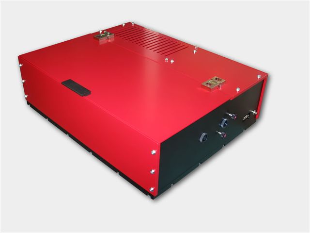
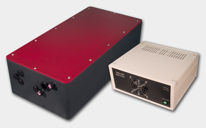
Tourmaline Yb-SS-1058/100 Femtosecond solid state laser system
The Yb-doped Tourmaline Yb-SS laser radiates at 1058±2 nm
with more than 1 W of average power, and enables the user to enjoy
Ti:Sapphire level power at over-micron wavelengths. This new design from Del
Mar's engineers features an integrated pump diode module for greater system
stability and turn-key operation. The solid bulk body of the laser ensures
maximum rigidity, while self-starting design provides for easy
"plug-and-play" operation.
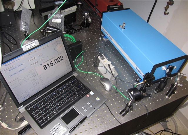
New laser spectrometer
T&D-scan for research that
demands high resolution and high spectral
density in UV-VIS-NIR spectral domains - now available with
new pump option! The T&D-scanincludes
a CW ultra-wide-tunable narrow-line laser, high-precision wavelength meter,
an electronic control unit driven through USB interface as well as a
software package. Novel advanced design of the fundamental laser component
implements efficient intra-cavity frequency doubling as well as provides a
state-of-the-art combined ultra-wide-tunable Ti:Sapphire & Dye laser
capable of covering together a
super-broad spectral range between 275 and 1100 nm. Wavelength
selection components as well as the position of the non-linear crystal are
precisely tuned by a closed-loop control
system, which incorporates highly accurate wavelength meter.
Near IR viewers
High performance infrared
monocular viewers are designed to observe radiation emitted by
infrared sources. They can be used to observe indirect radiation of IR
LED's and diode lasers, Nd:YAG, Ti:Sapphire, Cr:Forsterite, dye lasers and
other laser sources. IR viewers are ideal for applications involving the
alignment of infrared laser beams and of optical components in
near-infrared systems. Near IR viewers
sensitive to laser radiation up to 2000 nm.
The light weight, compact monocular may be used as a hand-held or facemask
mounted for hands free operation.
Ultraviolet viewers are
designed to observe radiation emitted by UV sources.
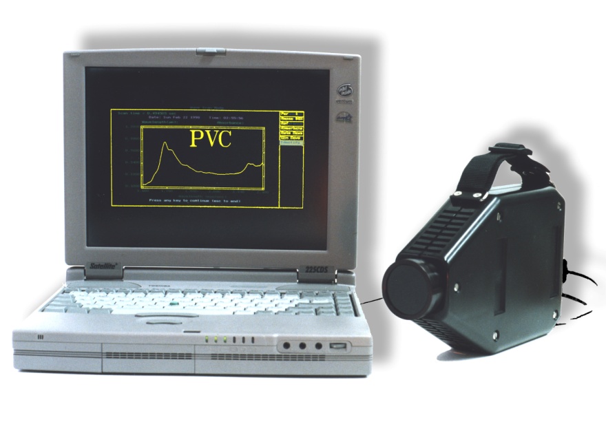
AOTF Infrared Spectrometer
Del Mar Photonics offer a handheld
infrared spectrometer based on the
acousto-optic tunable filter (AOTF). This instrument is about the size and
weight of a video camera, and can be battery operated. This unique, patented
device is all solid-state with no moving parts. It has been sold for a wide
variety of applications such as liquid fuel analysis, pharmaceutical analysis,
gas monitoring and
plastic analysis.
Miniature AOTF infrared spectrometer uses
a crystal of tellurium dioxide to scan the wavelength. Light from a light source enters
the crystal, and is diffracted into specific wavelengths. These wavelengths are
determined by the frequency of the electrical input to the crystal. Since there
are no moving parts, the wavelength scanning can be extremely fast. In addition,
specific wavelengths can be chosen by software according to the required algorithm, and therefore can be modified without changing the
hardware. After the infrared radiation reflects off of the sample, it is
converted into an electrical signal by the detector and analyzed by the
computer. Del Mar Photonics is looking for international distributors for
RAVEN - AOTF IR spectrometer for plastic identification and for variety of
scientific and industrial collaborations to explore futher commercial potential
of AOTF technology.
New:
AOTF spectrometer to measure lactose, fat and proteins in milk
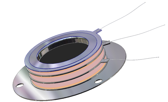
Open Microchannel Plate Detector
MCP-MA25/2 -
now in stock!
Microchannel Plate Detectors MCP-MA series are an open MCP detectors
with one or more microchannel plates and a single metal anode. They are intended
for time-resolved detection and make use of high-speed response properties of
the MCPs. MCP-MA detectors are designed for photons and particles detection in
vacuum chambers or in the space.
MCP-MA detectors are used in a variety of applications including UV, VUV and EUV
spectroscopy, atomic and molecular physics, TOF mass–spectrometry of clusters
and biomolecules, surface studies and space research.
MCP-MA detectors supplied as a totally assembled unit that can be easily mounted
on any support substrate or directly on a vacuum flange. They also can be
supplied premounted on a standard ConFlat flanges.
buy online -
ask for research discount!
Hummingbird EMCCD camera
The digital Hummingbird
EMCCD camera combines high sensitivity, speed and high resolution.
It uses Texas Instruments' 1MegaPixel Frame Transfer Impactron device which
provides QE up to 65%.
Hummingbird comes with a standard CameraLink output.
It is the smallest and most rugged 1MP EMCCD camera in the world.
It is ideally suited for any low imaging application such as hyperspectral
imaging, X-ray imaging, Astronomy and low light surveillance.
It is small, lightweight, low power and is therefore the ideal camera for
OEM and integrators.
buy online
Hatteras-D
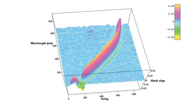
femtosecond transient absorption data acquisition system
Future nanostructures and biological nanosystems will take
advantage not only of the small dimensions of the objects but of the
specific way of interaction between nano-objects. The interactions
of building blocks within these nanosystems will be studied and optimized on
the
femtosecond time scale - says Sergey Egorov, President and CEO of Del Mar
Photonics, Inc. Thus we put a lot of our efforts and resources into the
development of new Ultrafast
Dynamics Tools such as our Femtosecond Transient Absorption Measurements
system Hatteras. Whether you want to
create a new photovoltaic system that will efficiently convert photon energy
in charge separation, or build a molecular complex that will dump photon energy
into local heat to kill cancer cells, or create a new fluorescent probe for
FRET microscopy, understanding of internal dynamics on femtosecond time scale
is utterly important and requires advanced measurement techniques.
Beacon Femtosecond Optically Gated Fluorescence Kinetic Measurement System
-
request a quote -
pdf
Beacon together with Trestles Ti:sapphire oscillator, second and third harmonic
generators. Femtosecond optical gating (FOG) method gives best temporal
resolution in light-induced fluorescence lifetime measurements. The resolution
is determined by a temporal width of femtosecond optical gate pulse and doesn't
depend on the detector response function. Sum frequency generation (also called
upconversion) in nonlinear optical crystal is used as a gating method in the
Beacon femtosecond fluorescence kinetic measurement system. We offer
Beacon-DX for operation together with Ti: sapphire femtosecond oscillators
and Beacon-DA for operation together with femtosecond amplified pulses.
Wavefront Sensors: ShaH Family
A family of ShaH wavefront sensors represents recent progress of Del Mar
Photonics in Shack-Hartmann-based technology. The performance of Shack-Hartmann
sensors greatly depends on the quality of the lenslet arrays used. Del Mar
Photonics. developed a proprietary process of lenslet manufacturing, ensuring
excellent quality of refractive lenslet arrays. The arrays can be AR coated on
both sides without interfering with the micro-lens surface accuracy. Another
advantage of the ShaH wavefront sensors is a highly optimized processing code.
This makes possible real-time processing of the sensor data at the rate
exceeding 1000 frames per second with a common PC. Due to utilizing low-level
programming of the video GPU, it is possible to output the wavefront data with a
resolution up to 512x512 pixels at a 500+ Hz frame rate. This mode is favorable
for controlling modern LCOS wavefront correctors.
The family of ShaH wavefront sensors includes several prototype models, starting
from low-cost ShaH-0620 suitable for teaching laboratory to a high-end
high-speed model, ShaH-03500. The latter utilizes a back-illuminated EM-gain CCD
sensor with cooling down to -100°C. This makes it possible to apply such a
wavefront sensor in astronomy, remote sensing, etc.
Fifth Harmonic Generator for
Nd:YAG lasers
The Fifth Harmonic Generator model LG105 is compatible with any pulsed
Nd:YAG laser, and is designed to produce UV-radiation at 213 nm. The
Nd:YAG laser, equipped with LG105, is a versatile device, and in many
applications can eliminate the necessity for excimer lasers. Solid state
technology that does not use toxic gases and costs less gives you the
advantages of both consistent, day-to-day operation and low maintenance. A
high quality BBO crystal is used in the LG105 as the non-linear element,
providing up to 20% conversion efficiency into 213 nm. The non-linear
crystal is placed in a special cell ensuring long lifetime of BBO without
any degradation or breakage. A harmonic separation system installed in LG105
provides nearly 100 % spectral purity of the output at 213 nm. The LG105
Fifth Harmonic Generator gives you not only high power output but also
excellent radiation stability
IntraStage lowers the cost
of test data management!
Struggling with gigabytes or terabytes of test data? IntraStage easily transforms test
data from disparate sources into web-based quality metrics and engineering
intelligence you can use.
Contact
us today to discuss your test management requirements and specifications of your
application.
Training Workshops
Come to San Diego next summer! Attend one of our training workshops in San Diego, California
during summer 2011
Del Mar Photonics has presented training workshops for
customers and potential customers in the past 3 years.
Our workshops cover scientific basics, technical details and provide
generous time for hands-on training.
Each workshop is a three-day seminar conducted by professional lecturer from 10am to 4pm. It includes lunch, as
well as a training materials. We have also reserved two days for Q&A sessions,
one-on-one system integration discussions, social networking, and San Diego sightseeing.
Del
Mar Photonics offers new
Trestles fs/CW laser system which can be easily
switched from femtosecond mode to CW and back. Having both modes of operation in one system dramatically increase a
number of applications that the laser can be used for, and makes it an ideal
tool for scientific lab involved in multiple research projects.
Kaelyn Leake is a PhD student in Electrical Engineering. She graduated from
Sweet Briar College with a B.S. in Engineering Sciences and Physics. Her
research interests include development of nanoscale optofluidic devices and
their applications. Kaelyn is the recipient of a first-year QB3 Fellowship.
In this video Kaelyn talks about her experimental research in nanoscale
optofluidics to be done with Trestles LH laser.
Frequency-stabilized CW
single-frequency ring Dye laser DYE-SF-007 pumped by DPSS DMPLH laser installed
in the brand new group of Dr. Dajun Wang at the The Chinese University of Hong
Kong.
DYE-SF-077 features exceptionally narrow generation line width, which
amounts to less than 100 kHz. DYE-SF-077 sets new standard for generation
line width of commercial lasers. Prior to this model, the narrowest line-width
of commercial dye lasers was as broad as 500 kHz - 1 MHz. It is necessary to
note that the 100-kHz line-width is achieved in DYE-SF-077 without the use of an
acousto-optical modulator, which, as a rule, complicates the design and
introduces additional losses. A specially designed ultra-fast PZT is used for
efficient suppression of radiation frequency fluctuations in a broad frequency
range. DYE-SF-077 will be used in resaerch of Ultracold polar molecules,
Bose-Einstein condensate and quantum degenerate Fermi gas and High resolution
spectroscopy
Del Mar Photonics continuously expands its components
portfolio.
Solar
Prisms for Concentrating Photovoltaic Systems (CPV) Solar cells made of compound semiconductors such
as gallium arsenide are very expensive. Usually very small cells are
installed and various means such as mirrors, lenses, prisms, etc..are used
to concentrate sunlight on the cells. Concentration photovoltaic technology
(CPV) uses the solar radiation with an efficiency of 40%, double that of
conventional solar cells
Del Mar Photonics design custom Concentrating Photovoltaic Systems (CPV) and
supply variety of the optical components for CPV such as
solar prisms shown in the picture.
Axicon Lens
Axicon lens also known as conical lens or rotationally symmetric prism is
widely used in different scientific research and application. Axicon can be
used to convert a parallel laser beam into a ring, to create a non
diffractive Bessel beam or to focus a parallel beam into long focus depth.
Del Mar Photonics supplies axicons with cone angles range from 130° to
179.5° for use with virtually any laser radiation. We manufacture and supply
axicons made from BK7 glass, fused silica and other materials.
Rutile (TiO2) coupling
prisms
Del Mar Photonics offers optical elements made of high quality synthetically
grown Rutile Titanium Dioxide crystals. Rutile’s strong birefringency, wide
transmission range and good mechanical properties make it suitable for
fabrication of polarizing cubes, prisms and optical isolators. Boules having
high optical transmission and homogeneity are grown by proprietary method.
Typical boules have 10 - 15 mm in dia. and up to 25 mm length. Optical
elements sizes - from 2 x 2 x 1 mm to 12.7 x 12.7 x 12.7 mm. Laser grade
polish quality is available for finished elements. So far we the largest
elements that we manufactured are 12 x15 x 5 mm, in which optical axis is
parallel to 15 mm edge, 5 mm is along beam path, 12 x 15 mm faces polished
20/10 S/D, one wave flatness, parallelism < 3 arc.min. (better specs.
available on request).
Vacuum viewport
Del Mar Photonics offer a range of competitively priced UHV viewports ,
Conflat, ISO or KF including a variety of coatings to enhance performance.
Del Mar Photonics viewports are manufactured using advanced techniques for
control of special and critical processes, including 100 percent helium leak
testing and x-ray measurements for metallization control. Windows Materials
include: Fused silica, Quartz , Sapphire , MgF2, BaF2, CaF2, ZnSe, ZnS, Ge,
Si, Pyrex. Standard Viewing diameters from .55" to 1.94 ".
Coating - a range of custom coatings can applied - which include
- Single QWOT
- Broad Band AR
- V coatings
- ITO
- DLC (Diamond like coating)
NARROW-BAND HOLOGRAPHIC FILTERS are intended for
suppression of powerful beams in research and in engineering, in particular, in laser
spectroscopy, and also for protection from blinding and damaging by laser radiation various photo
receiver devices and operator's eyes.
Unlike conventional interference filters, which are made by vacuum
evaporation techniques, holographic filters are fabricated by recording
interference patterns formed between two mutually coherent laser beams. Since all layers are recorded simultaneously within a thick stack, the
optical density of the notch filter is high and its spectral bandwidth
can be extremely narrow. Also, since the layering profile is sinusoidal
instead of square wave, holographic notch filters are free from
extraneous reflection bands and provide significantly higher laser
damage thresholds.
Hydrogen
Thyratrons are used in
such devices as radars with different power levels, high-power pulsed
technical, electrophysical, medical devices and lasers. Sophisticated
design and high quality ceramic-metal envelope determines long lifetime
and very accurate and reliable operation of hydrogen thyratrons under wide range of environmental
conditions. Applications:
- radars
- pulsed lasers power supplies
- medical apparatus
- electrophysical instrumentation
Triggered Three-Electrode Spark Gap Switches are ceramic-metal sealed off gas
discharge trigatron-type devices with a co-axial trigger electrode. These Gas
Discharge Tubes contain no mercury and, due to an advanced design, feature high
reliability and a long lifetime being operating under wide range of
environmental conditions. Applications:
- pulsed installation for processing materials
- installations with plasma focus
- pulse power supplies for lasers and other pulse equipment
- medical apparatus such as lithotriptors and defibrillators
- processing systems for petroleum wells
Trigger Transformers
Del Mar Photonics supply
trigger transformers for triggered spark gaps and
other applications.
Contact us to today to discuss your application or requesta quote.
Trigger Transformers are used to provide a fast high voltage pulse up to
30kV/µs and more. This high voltage pulse is applied to the trigger
electrode to initiate switching action in the
three-electrode
spark gaps. Either positive or negative pulses can be obtained from all
of the transformers.
We are looking forward to hear from you and help you with
your optical and crystal components requirements. Need time to think about
it?
Drop us a line and we'll send you beautiful Del Mar Photonics mug (or
two) so you can have a tea party with your colleagues and discuss your
potential needs.
Del Mar Photonics, Inc. 4119 Twilight Ridge
San Diego, CA 92130
tel: (858) 876-3133
fax: (858) 630-2376
Skype: delmarphotonics sales@dmphotonics.com